Comprehensive Guide to Office Hysteroscopy: A Safe and Effective Gynecological Procedure
In the realm of modern gynecology, office hysteroscopy has emerged as a groundbreaking procedure, transforming the way healthcare providers diagnose and treat a variety of uterine conditions. This minimally invasive technique offers numerous advantages over traditional surgical approaches, putting patient comfort, safety, and rapid recovery at the forefront of women’s reproductive health.
Understanding Office Hysteroscopy: What Is It?
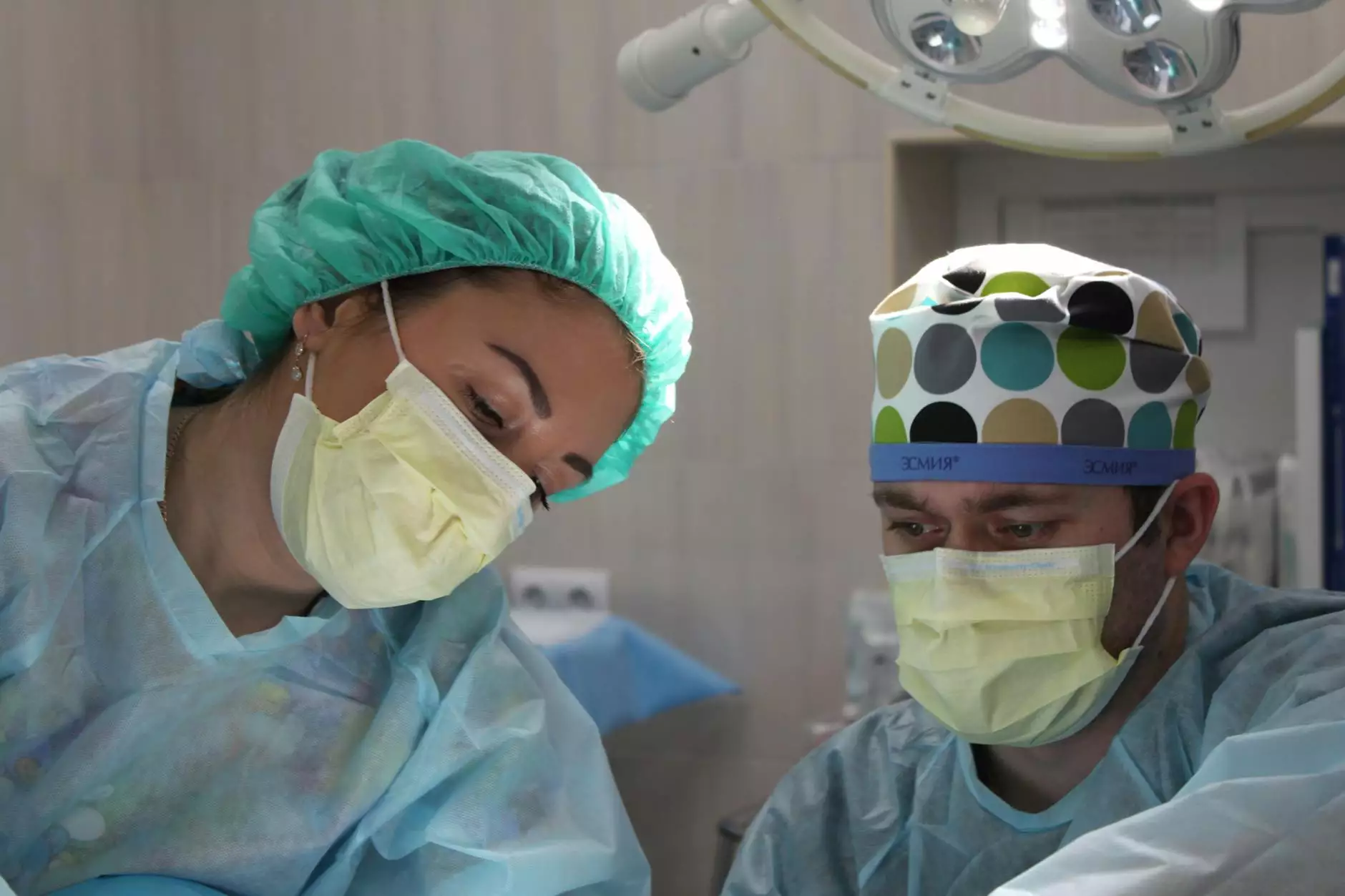
At its core, office hysteroscopy is a diagnostic and therapeutic procedure that allows gynecologists to directly visualize the inside of a woman's uterine cavity using a thin, lighted telescope called a hysteroscope. Unlike traditional hysteroscopy performed in an operating room, office hysteroscopy is conducted in a clinical setting, often without general anesthesia, making it more accessible and less intimidating for women.
The Evolution of Hysteroscopic Technology in Modern Gynecology
Historically, hysteroscopic procedures were confined to inpatient settings, entailing significant discomfort and requiring anesthesia. However, advancements in endoscopic technology and miniaturization of instruments have expanded the applicability of hysteroscopy into outpatient care.
Modern office hysteroscopy uses high-definition cameras, ultra-fine rigid or flexible hysteroscopes, and specialized tools that enable precise diagnosis and targeted treatment during a single visit. This technological leap has positioned office hysteroscopy as a critical component of minimally invasive gynecology.
Key Indications for Office Hysteroscopy
This procedure is indicated for a variety of gynecological concerns, including:
- Menstrual irregularities: Abnormal bleeding patterns, menorrhagia, and intermenstrual bleeding
- Reproductive issues: Unexplained infertility and recurrent miscarriage investigations
- Follow-up procedures: Post-ablation assessment or removal of intrauterine devices
- Uterine abnormalities: Polyps, fibroids, septa, and adhesion assessments and removals
- Assessment of intrauterine pathology: To diagnose causes of abnormal uterine bleeding or suspected lesions
Advantages of Office Hysteroscopy
The shift towards outpatient, office-based procedures introduces several significant benefits:
- Minimally invasive: No need for incisions or hospital stay, resulting in less trauma and faster recovery.
- High precision and visualization: Direct real-time visualization facilitates accurate diagnosis and targeted treatment.
- Patient comfort: Procedures are typically performed without general anesthesia, reducing risks and anxiety.
- Cost-effectiveness: Lower overall healthcare costs due to reduced hospital stays and anesthesia requirements.
- Convenience: Short outpatient visits with minimal downtime enable women to return to daily activities swiftly.
Pre-Procedure Preparation for Office Hysteroscopy
Proper preparation enhances the safety and effectiveness of the procedure. It generally involves:
- Thorough consultation with the gynecologist to discuss medical history and any medications
- Potential use of pre-procedure medications such as NSAIDs to minimize cramping
- Timing the procedure during the proliferative phase of the menstrual cycle for optimal visualization
- Informing the patient about what to expect to alleviate anxiety and ensure compliance
- Ensuring clearance of any menstrual bleeding, as this could interfere with visibility
The Step-by-Step Process of Office Hysteroscopy
While specific techniques may vary slightly depending on the clinical setting, the typical steps are as follows:
1. Patient Positioning and Anesthesia
The patient is positioned in lithotomy, and local anesthesia or a vasoconstrictive agent (such as diluted epinephrine) might be applied to the cervical canal to reduce discomfort. Often, no general anesthesia is required, making the procedure less burdensome.
2. Cervical Dilation and Insertion of Hysteroscope
The clinician gently dilates the cervix if necessary, then introduces the hysteroscope before insufflating the uterine cavity with a small amount of sterile saline or carbon dioxide gas for visualization.
3. Visual Examination and Identification of Pathology
The high-definition camera provides real-time images, allowing the practitioner to carefully examine the endometrial lining and identify any anomalies such as polyps, fibroids, or adhesions.
4. Targeted Treatment
If abnormalities are detected, the hysteroscope is used to perform interventions like polyp removal, septum resection, or fibroid excision, often with integrated surgical tools. This all occurs during the same session, eliminating the need for additional surgeries.
5. Completion and Post-Procedure Care
Once the procedure is complete, the hysteroscope is withdrawn, and the patient is monitored briefly for any immediate adverse reactions. Most women experience minimal discomfort and can resume normal activities shortly afterward.
Safety Considerations and Risks of Office Hysteroscopy
Thanks to technological advances and improved techniques, office hysteroscopy is generally regarded as a very safe procedure. Nonetheless, some risks include:
- Uterine perforation: Rare but possible, especially if the uterus is enlarged or have abnormal anatomy
- Infection: Usually preventable with proper sterile techniques
- Vasovagal responses or dizziness: Typically mild and manageable
- Cramping or discomfort: Usually transient and alleviated with analgesics
Adherence to strict procedural protocols and patient selection ensures minimal complication rates, making it a reliable option for appropriate candidates.
Post-Procedure Expectations and Follow-Up
Following office hysteroscopy, women may experience mild cramping, light bleeding, or discharge. These symptoms are typically self-limited. It is advisable to:
- Relax and rest as needed
- Use prescribed analgesics if discomfort occurs
- Monitor for signs of infection, such as fever or foul discharge
- Follow up with the healthcare provider for results and further recommendations
In cases where surgical removal of polyps or fibroids was performed, additional treatments or imaging might be necessary to confirm the success of intervention.
The Role of Office Hysteroscopy in Women's Reproductive Health
The ability to diagnose and treat intrauterine conditions in a minimally invasive, outpatient setting has significantly enhanced women's healthcare. Office hysteroscopy plays a pivotal role in:
- Improving fertility outcomes: Addressing intrauterine abnormalities that hinder conception
- Managing abnormal bleeding: Providing definitive diagnosis and treatment
- Reducing the need for invasive surgeries: Avoiding complications and lengthy recoveries
- Empowering women: Enabling greater participation in reproductive health decisions
Choosing the Right Specialist for Your Office Hysteroscopy
Given the precision required, selecting a highly experienced Gynecologist, preferably with specialized training in minimally invasive procedures, is paramount. Dr. Seckin and his team at drseckin.com are renowned for their expertise in reproductive health and gynecological endoscopic procedures. Their commitment to patient-centered care ensures optimal outcomes and comfort during your office hysteroscopy.
Final Thoughts: Embracing Modern Gynecological Care with Office Hysteroscopy
The continuous evolution of gynecological techniques underscores the importance of minimally invasive, outpatient procedures for women's health. Office hysteroscopy embodies this progress, offering an effective, safe, and patient-friendly solution to diagnose and treat a wide array of uterine conditions. Its integration into routine reproductive care signifies a new era where women can access high-quality healthcare with minimal disruption to their lives.
By understanding the benefits, process, and safety profile of office hysteroscopy, women can make informed decisions about their reproductive health and seek expert care to improve their overall well-being.